Dental Radiology
Dental Radiology provide the basis for the complete and accurate diagnosis of almost every dental condition.
Dr. Zafer Kazak and the Medicadent Team are dedicated to providing you with the highest standard of care by using only the best technology available, in the most sterile environment to benefit your health.
Digital volume tomography (DVT) provides an opportunity to depict the bony structures of the jaw three dimensionally. The great advantage compared to computed tomography (CT) is a significantly lower radiation exposure.
With the help of this image, surgical procedures can be planned in advance if needed and thus be performed much more gentle and safe. This is particularly the case for implants.
We have such a device in our surgery clinic so that, if required, an image can be produced straightaway.
Benefits of digital dental radiographs compared to traditional dental X-rays include the following:
Digital radiographs reveal small hidden areas of decay between teeth or below existing restorations (fillings), bone infections, gum (periodontal) disease, abscesses or cysts, developmental abnormalities and tumors that cannot be detected with only a visual dental examination.
Digital radiographs can be viewed instantly on any computer screen, manipulated to enhance contrast and detail, and transmitted electronically to specialists without quality loss.
Early detection and treatment of dental problems can save time, money and discomfort.
Digital micro-storage technology allows greater data storage capacity on small, space-saving drives.
Dental digital radiographs eliminate chemical processing and disposal of hazardous wastes and lead foil, thereby presenting a “greener” and eco-friendly alternative.
Digital radiographs can be transferred easily to other dentists with compatible computer technology, or photo printed for dentists without compatible technology.
Digital dental images can be stored easily in electronic patient records and, sent quickly electronically to insurance companies, referring dentists or consultants, often eliminating or reducing treatment disruption and leading to faster dental insurance reimbursements.
How often the x-rays are needed depends on the medical and dental history and current condition of the patient. Some people need the x-rays every six months. For some people x-rays can be needed in a period of two years.
• Children: Depending on age, the x-rays are needed for the most of the children every six months to one year. Because the decay can be formed easily in the children’s mouth. By the help of the x-rays, it is possible to monitor the tooth development too.
• Adults with extensive restoration work: It is important for the adults with extensive restoration work, including fillings has to be taken regularly x-rays. Because the risk of the problem formation in these mouths increases much more faster. Anyone who drinks sugary sodas, chocolate milk or coffee or tea with sugar
• People with periodontal (gum) disease: The people who have the periodontal disease and who are having a periodontal treatment may need the x-rays more often in order to see if there are significant or continuing signs of bone loss.
• People who are taking medications that lead to dry mouth: In a dry mouth because of the lost of the saliva the tooth decays may be formed easily because saliva helps to keep the acid levels in the mouth stable and when the saliva degree decreases, the acid levels increases and that cause the tooth decay. Some medications cause the dry mouth. The medications prescribed for hypertension, antidepressants, antianxiety drugs, antihistamines, diuretics, narcotics, anticonvulsants and anticholinergics are between the medications which cause the dry mouth. The people who have the dry mouth problem may need the x-rays more often.
• Smokers: The smokers need the x-rays more often because smoking increases the risk of periodontal disease.
- Panoramic Radiographs
- Tomogram
- Computed tomography or Ct scanning
Digital radiographs are one of the newest X-ray techniques around. It reduces radiation by as much as 80 percent. With digital radiographs, film is replaced with a flat electronic pad or sensor. The X-rays hit the pad the same way they hit the film.
But instead of developing the film in a dark room, the image is electronically sent directly to a computer where the image appears on the screen. The image can then be stored on the computer or printed out
Benefits of digital dental radiographs compared to traditional dental X-rays include the following:
- Digital radiographs reveal small hidden areas of decay between teeth or below existing restorations (fillings), bone infections, gum (periodontal) disease, abscesses or cysts, developmental abnormalities and tumors that cannot be detected with only a visual dental examination.
- Digital radiographs can be viewed instantly on any computer screen, manipulated to enhance contrast and detail, and transmitted electronically to specialists without quality loss.
- Early detection and treatment of dental problems can save time, money and discomfort.
- Digital micro-storage technology allows greater data storage capacity on small, space-saving drives.
- Dental digital radiographs eliminate chemical processing and disposal of hazardous wastes and lead foil, thereby presenting a “greener” and eco-friendly alternative.
- Digital radiographs can be transferred easily to other dentists with compatible computer technology, or photo printed for dentists without compatible technology.
- Digital dental images can be stored easily in electronic patient records and, sent quickly electronically to insurance companies, referring dentists or consultants, often eliminating or reducing treatment disruption and leading to faster dental insurance reimbursements
Laser Treatment
Dentists use lasers for more than 20 Years to treat a number of dental problems. These lasers are different from the cold lasers used in other fields. State-of-the-art laser treatment makes for a significantly more pleasant experience than previously possible.
Our clinic is equipped with the latest laser technology and is the partner of “AALZ-Aachener Laser Centre” in Germany. We are training clinic in Turkey for the laser manufacturer Fotona and in addition, external dentists get training continuously in dealing with the laser by Dr. Kazak, founder of Medicadent.
The laser can be used in a number of different ways, i.e.
· Infected tissue can be vaporised using laser, without damaging the tooth enamel
· Necks of teeth that are sensitive to cold / hot can be “sealed” without any pain at all
· Herpes or mouth ulcers heal faster if radiated gently.
· Targeted disinfection and germ reduction for treating gum diseases or root canal treatments
· Optimises the success of the overall treatment
· The tiny grooves in children’s teeth can be disinfected without “drilling” before they are sealed
· Thorough removal of any remaining caries
· Preserve any teeth that are at risk
· There is no need for scalpels or stiches because the laser closes any blood vessels immediately
Here are some of the major benefits associated with laser dentistry:
- Procedures performed using soft tissue dental lasers may not require sutures (stitches).
- Certain laser dentistry procedures do not require anesthesia.
- Laser dentistry minimizes bleeding because the high-energy light beam aids in the clotting (coagulation) of exposed blood vessels, thus inhibiting blood loss.
- Bacterial infections are minimized because the high-energy beam sterilizes the area being worked on.
- Damage to surrounding tissue is minimized.
- Wounds heal faster and tissues can be regenerated.
The Food and Drug Administration (FDA) has approved of a variety of hard and soft tissue lasers for use in the dental treatment of adults and children. Because dental lasers boast unique absorption characteristics, they are used to perform specific dental procedures.
Hard Tissue Lasers: Hard tissue lasers have a wavelength that is highly absorbable by hydroxyapatite (calcium phosphate salt found in bone and teeth) and water, making them more effective for cutting through tooth structure. Hard tissue lasers include the Erbium YAG and the Erbium chromium YSGG. The primary use of hard tissue lasers is to cut into bone and teeth with extreme precision. Hard tissue lasers are often used in the “prepping” or “shaping” of teeth for composite bonding, the removal of small amounts of tooth structure and the repair of certain worn down dental fillings.
Soft Tissue Lasers: Soft tissue lasers boast a wavelength that is highly absorbable by water and hemoglobin (oxygenating protein in red blood cells), making them more effective for soft tissue management. Commonly used soft tissue lasers include Neodymium YAG (Nd:YAG) and diode lasers, which may be used as a component of periodontal treatment and have the ability to kill bacteria and activate the re-growth of tissues. The carbon-dioxide laser minimizes damage to surrounding tissue and removes tissue faster than the fiber optic method. Soft tissue lasers penetrate soft tissue while sealing blood vessels and nerve endings. This is the primary reason why many people experience virtually no postoperative pain following the use of a laser. Also, soft tissue lasers allow tissues to heal faster. It is for this reason that a growing number of cosmetic dental practices are incorporating the use of soft tissue lasers for gingival sculpting procedures.
Some dental laser technology has been developed that can be used to generate both hard and soft tissue laser energy, depending upon the patient’s needs.
In addition to the lasers used for cutting and shaping hard and soft tissues, other laser types are specifically designed for viewing the insides of teeth and cells using Optical Coherence Tomography, a non-invasive imaging technique. Other lasers provide energy and specific proteins that help move messages between cells to match the body’s natural ability to use light spectrums to heal damaged cells.
Consult Medicadent for more information about the types and benefits of procedures performed in laser dentistry.
If the dental laser is at least as safe as other dental instruments. However, just as you wear sunglasses to protect your eyes from prolonged exposure to the sun, when your dentist performs a laser procedure, you will be asked to wear special eyeglasses to protect your eyes from the laser.
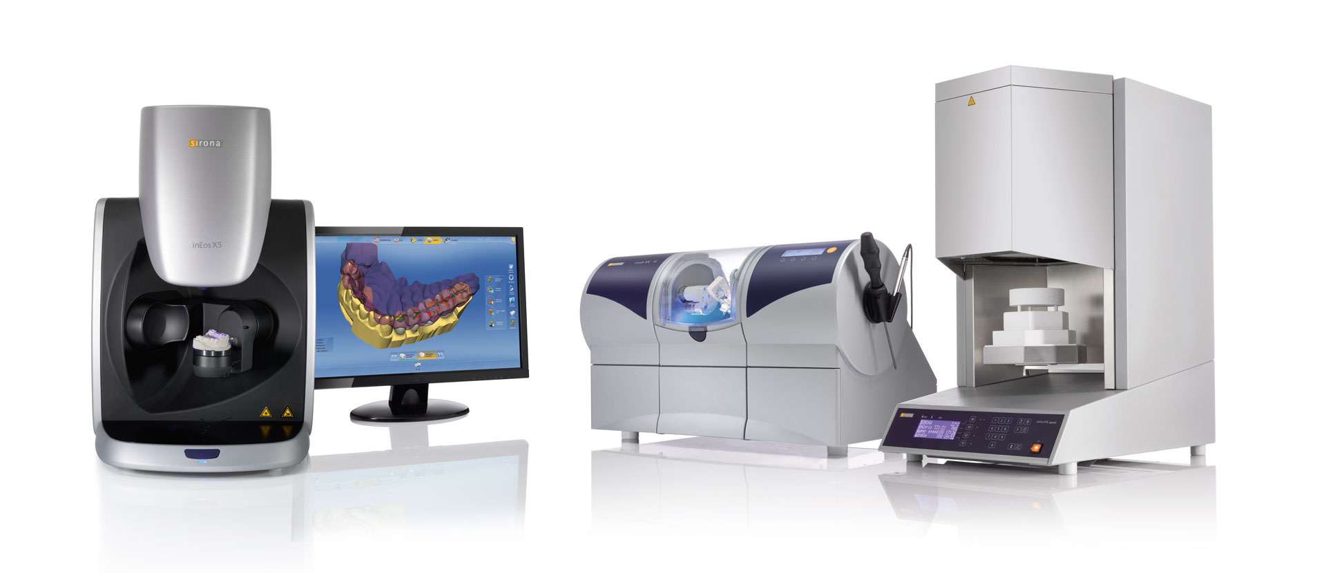
CAD/CAM (Computer aided design and computer aided manufacturing)

Digital solution
Modern dentistry is different from traditional dentistry, where treatment is quick to maintain patient comfort and provide the best quality for the patient.
Through this advanced technology , it is possible to do a dental examination and after scanning the teeth can print the image . and in a very short time, less pain and more effective way we can provide cosmetic solutions to the missing teeth in the patient’s mouth.
The digital impressions
Digital impressions allows dentists to create a virtual, computer-generated replica of the hard and soft tissues in the mouth using lasers and other optical scanning devices. The digital technology captures clear and highly accurate impression data in few minutes, without the need for traditional impression materials that some patients find inconvenient and messy. Many patients find digital impressions an easier and more comfortable procedure because traditional impression materials are avoided. The impression information then is transferred to a computer and used to create restorations.
Do you have a problem with the long period of treatment for your teeth and the time spent in visiting your doctor?
Digital impressions can be used to make same day dentistry restorations, thereby speeding up patient treatment and reducing the need for multiple clinic visits.
Do you want to see the future smile that will be presented to you at the end of the treatment?
In the cases which the patient needs to beautify the shape of his smile, after we make a scan for the hard and soft tissues in the mouth , and by modern programs we can prepare the perfect smile design for the condition of this patient while he is in the clinic.
The device will simulate the situation and extract the appearance that will be the result of the case cosmetic solution and the program allows the possibility of the modification of this result by the patient and the doctor together.
Do you want to eliminate the fear of dental implants and minimize their surgical risk?
A dental implant is designed to replace the root part of the tooth. To replace the visible part of the tooth, a crown, bridge or denture can be attached once the implant is secure , which may be the same day or several weeks later, depending on the individual situation. Dental implants are made of titanium .
Before placing the implant, a lot of planning goes on — typically involving X-rays , and sometimes CT scans. This ensures that the operation itself goes smoothly. When it’s time for the procedure you’ll receive a local anesthetic, and we’ll make sure you don’t feel anything.
Do you find the idea of a mouthful of metal braces and brackets – however effective – too unattractive to commit to?
As an alternative to more traditional orthodontic solutions, which entail metal braces and brackets, Medicadent Invisalign offers an orthodontic adjustment method that uses a series of clear aligners, or trays, to gradually adjust your teeth and alignment.
The apparatus is clear, almost invisible and is a comfortable, effective, aesthetically pleasing way to get a straight and healthy mouth.